Anwer Khan Modern Medical College

Library Management System
Advance Search

Book Detail

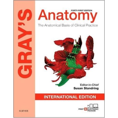
Title: Gray's anatomy: the anatomical basis of clinical practice
Author Name: Susan Standrings [Editor in Chief].
Author Sur Name: STANDRINGS, Susan.
Author information:
Emeritus Professor of Anatomy at King’s College London, Susan Standring, MBE, PhD, DSc, Hon FRCS, is a neuroscientist who worked on the biology of repair in the peripheral nervous system for many years. She is a Past President of the Peripheral Nerve Society. As an anatomist, she taught medical and dental students and surgical trainees for 40 years and has championed anatomy as a fundamental component of medical and dental curricula throughout her career. Professor Standring a Past President of the Anatomical Society and Deputy Chair of the Board of Trustees of the Hunterian Collection at the Royal College of Surgeons of England. She is a Fellow of King’s College London and an Honorary Fellow of RCSEng. She was awarded the prestigious Wood Jones Medal of RCSEng for her outstanding contribution to anatomy teaching.
Edition/Published: 41st ed. _New Delhi : Elsevier , 2016
New to this edition:
  1. Each chapter has been edited by experts in their field, ensuring access to the very latest evidence-based information on that topic.
  2. More than 1,000 completely new photographs, including an extensive electronic collection of the latest X-ray, CT, MR, and histological images.
  3. The downloadable Expert Consult eBook version included with your purchase allows you to search all of the text, figures, references and videos from the book on a variety of devices.
  4. Carefully selected electronic enhancements include additional text, tables, illustrations, labelled imaging and videos – as well as 24 specially invited ‘Commentaries’ on new and emerging topics related to anatomy.
Physical Description:
xviii, 1562, : illustrations (chiefly color), tables, graphs.; 30cm.
Notes Includes Bibliographical References and Indexes.
Includes Index: P. 1453-1562
ISBN No's: 978-0-7020-6306-0 ,
Bar Code's: ,
Shelf Location's: 98 ,
Classification
Subject: Human Anatomy
Dewey Class No: 611
Letter Call No: S1g
LC Classification: QM23.2 .G73 2016
Other's Book Information
Book ID No: 1658 , 1659
Total Books: 2
Date of collection's: 01-Nov-2016 , 01-Nov-2016
Donation / Purchase: Purchase
Language: English
Status: Available
Department: Anatomy
Synopsis:
  1. In 1858, Drs. Henry Gray and Henry Vandyke Carter created a book for their surgical colleagues that established an enduring standard among anatomical texts. After more than 150 years of continuous publication, Gray’s Anatomy remains the definitive, comprehensive reference on the subject, offering ready access to the information you need to ensure safe, effective practice. This 41st edition has been meticulously revised and updated throughout, reflecting the very latest understanding of clinical anatomy from field leaders around the world. The book’s traditional lavish art programme and clear text have been further honed and enhanced, while major advances in imaging techniques and the new insights they bring are fully captured in new state-of-the-art X-ray, CT, MR, and ultrasonic images. The completely reconfigured accompanying eBook version is richly enhanced with additional content and media, covering all the body regions, cell biology and embryogenesis. This unlocks a whole new level of related information, interactivity and understanding, in keeping with the spirit of innovation that has characterized Gray’s Anatomy since its inception.
Review:
  1. "The newest (2015) and the 41st edition of Gray’s Anatomy is a mirror on the explosion of medical knowledge that has occurred over the past 8 years since the 40th edition was published… the 41st edition of Gray’s Anatomy is the single most important text a radiologist should own. Buy it not only for its practical and intellectual/educational value but to have in your possession-a slice of medical history." -American Journal of Neuroradiology (March 2016)
  2. "This edition of Gray's has gone a long way to being the perfect anatomy text. It has beautifully clear anatomical diagrams.The text is well edited; it is clear, concise and readable." British Journal of Oral and Maxillofacial Surgery, Volume 47(4), June 2009
Description:
  1. Gray's Anatomy 41st edition reflects the very latest understanding of clinical anatomy from field leaders worldwide. Its traditional artwork and clear text has been further honed and enhanced, while major advances in imaging techniques and the newest insights they bring are fully captured in new state-of-the-art x-ray, CT, MR and ultrasound images. The completely reconfigured accompanying eBook version is enhanced with additional content and media, covering all the body regions, cell biology and embryogenesis.
Key Features:
  1. Presents the most detailed and dependable coverage of anatomy available anywhere.
  2. Regional organization collects all relevant material on each body area together in one place, making access to core information easier for clinical readers.
  3. Anatomical information is matched with key clinical information where relevant.
  4. Numerous clinical discussions emphasize considerations that may affect medical care.
  5. Gray’s anatomy receives published under the banner of very famous Churchill Livingstone that’s a evidence of reliability and trust.
  6. You can understand each and everything present in the book with the assist of diagrams and 3-D snap shots which might be a incredible component of the book.
  7. Gray’s anatomy is one of the oldest anatomy book.
  8. Gray’s anatomy has 41 edition and frequently updated.
  9. It has an electronic form that is much more detailed. You can get free online access with the hard copy of this book.
  10. Electron microscope and florescence microscope diagrams are added. All the diagrams including both anatomical and microscopic diagrams are of great quality and you don’t need any anatomy atlas like netter atlas or grant’s atlas to use along with this anatomy book.
  11. Not only clinical gross anatomy but topics of embryology and histology are also touches along with the diagrams.
  12. This is the ideal anatomy book for students, anatomist, clinicians and teachers.
  13. References are given at the end of each chapter.
  14. You can easily find topic of your need at the end of book from index.
Summary:
"The 41st edition of Gray's Anatomy provides an interactive experience to engage users like never before," said Linda Belfus, Senior Vice President of Content, Elsevier Clinical Solutions. "After more than 150 years of continuous publication, Gray's Anatomy remains the definitive and most comprehensive anatomy reference that can help ensure safe and effective practice, enabling healthcare practitioners to offer their patients the best possible medical care."
Abstract:
41st edition of Gray's Anatomy: The Anatomical Basis of Clinical Practice. This latest edition of the renowned anatomical reference text used by medical practitioners and students alike has been meticulously revised and updated by a world-leading group of clinicians and anatomists building on more than 150 years of anatomical knowledge.
Preface:
‘Anatomy is the basis of medical discourse.’ (Hippocrates, De locis in homine 2) Looking through an almost complete set of the previous editions of Gray’s Anatomy, I am struck by the marked difference in size between the first and fortieth editions. That progressive increase in girth has occurred pari passu with ground-breaking advances in basic science and clinical medicine over the past 155 years. Anatomy has become a far wider discipline than Henry Gray, Henry van Dyke Carter or any of their students could have envisaged. Fields such as cell biology, molecular genetics, neuroanatomy, embryology and bioinformatics either had not emerged or were in their infancy in 1858. Techniques that today inform our view of the internal landscape of the body – such as specialized types of light and electron microscopy; imaging modalities, including X-rays, magnetic resonance imaging, computed tomography and ultrasonography; the use of ‘soft’ perfusion techniques and frozen-thawed, unembalmed cadavers for dissection-based studies; and the advances in information technology that enable endoscopic and robotic surgery and facilitate minimally invasive access to structures previously considered inaccessible – were all unknown. As each development entered mainstream scientific or clinical use, the new perspectives on the body it afforded, whether at submicroscopic or macroscopic level, filtered into the pages of Gray’s Anatomy: for example, the introduction of X-ray plates (twenty-seventh edition, 1938) and electron micrographs (thirtysecond edition, 1958). In the Preface to the first edition, Henry Gray wrote that ‘This Work is intended to furnish the Student and Practitioner with an accurate view of the Anatomy of the Human Body, and more especially the application of this science to Practical Surgery.’ We remain true to his intention. An appropriate knowledge of clinically relevant, evidence-based anatomy is an essential element in the armamentarium of a practising clinician; indeed, ‘If anything, the relevance of anatomy in surgery is more important now than at any other time in the past’ (Tubbs, in Preface Commentary, which accompanies this volume). In my Preface to the fortieth edition, I intimated that the book was quite literally in danger of breaking its binding if any more pages were added. In order to avoid this unfortunate occurrence, the forty-first edition contains a significant amount of material that is exclusively electronic, in the form of 77,000 words of additional text, 300 artworks and tables, 28 videos and 24 specially invited commentaries on topics as diverse as electron microscopy and fluorescence microscopy; the neurovascular bundles of the prostate; stem cells in regenerative medicine; the anatomy of facial ageing; and technical aspects and applications of diagnostic radiology. In keeping with the expectation that anatomy should be evidence-based, the forty-first edition contains many more references in the e-book than could be included in the thirty-ninth and fortieth printed editions. Neel Anand, Rolfe Birch, Pat Collins, Alan Crossman, Michael Gleeson, Ariana Smith, Jonathan Spratt, Mark Stringer, Shane Tubbs, Alan Wein and Caroline Wigley brought a wealth of scholarship and experience as anatomists, cell biologists and clinicians to their roles as Section Editors. I thank them for their dedication and enthusiastic support, in selecting and interacting with the authors in their Sections and for meeting deadlines, despite the ever-increasing demands on their time from university and/or hospital managers. Pat Collins, Girish Jawaheer, Richard Tunstall and Caroline Wigley worked closely with many authors to update the text and artworks for organogenesis, paediatric anatomy, evidence-based surface anatomy and microstructure, respectively, across Sections 3 to 9. Jonathan Spratt acted as both a Section Editor (thorax) and an indefatigable ‘go to’ for sourcing images throughout the book; in the latter capacity, he has produced a superb collection of additional labelled images, available in the e-book (see Bonus imaging collection). Over a hundred highly experienced anatomists and clinicians contributed text, often extensively revised from the previous edition, and/or artworks, original micrographs or other images to individual chapters. As a general rule, the orientation of diagrams and photographs throughout the book has been standardized to show the left side of the body, irrespective of whether a lateral or medial view is presented, and transverse sections are viewed from below to facilitate comparison with clinical images. Clinicopathological examples have been selected where the pathology is either a direct result, or a consequence, of the anatomy, or where the anatomical features are instrumental in the diagnosis/ treatment/management of the condition. Wherever possible, the photomicrographs illustrate human histology and embryology; non-human sources are acknowledged in the captions. In an ideal world, anatomical terminology would satisfy both anatomists and clinicians. For the avoidance of doubt, the same word should be agreed and used for each structure that is described, whether in the anatomy laboratory or the clinic. In the real world, this goal is achieved with varying degrees of success; alternative terms (co) exist and may (and frequently do) confuse or frustrate. Currently, Terminologia Anatomica (TA)1 is the reference source for the terminology for macroscopic anatomy; the text of the forty-first edition of Gray’s Anatomy is almost entirely TA-compliant. However, where terminology is at variance with, or, more likely, is not included in, the TA, the alternative term that is chosen either is cited in the relevant consensus document or position paper – e.g. ‘European Position Paper on the Anatomical Terminology of the Internal Nose and Paranasal Sinuses’2 and the International Interdisciplinary Consensus Statement on the ‘Nomenclature of the Veins of the Lower Limbs’3 – or enjoys widespread clinical usage: for example, the use of attitudinally appropriate terms in cardiology (see Chapter 57). The continued use of eponyms is contentious.4 Proponents of their retention argue that some eponyms are entrenched in medical language and are (therefore) indispensable, that they facilitate communication because their use is so pervasive and that they serve to remind us of the humanism of medicine. Detractors argue that eponyms are inherently inaccurate, non-scientific and often undeserved. In this edition of Gray’s Anatomy, synonyms and eponyms are given in parentheses on first usage of a preferred term and not shown thereafter in the text; an updated list of eponyms remains available in the e-book for reference purposes. I offer my sincere thanks to the editorial team at Elsevier, initially under the leadership of Madelene Hyde and latterly of Jeremy Bowes, for their guidance, professionalism, good humour and unfailing support. In particular, I thank Poppy Garraway, Humayra Rahman Khan, Wendy Lee, Joanna Souch, Julie Taylor, Jan Ross and Louise Cook, for being at the end of a phone or available by e-mail whenever I needed advice or support. I dedicate my work on the forty-first edition of Gray’s Anatomy to the memory of my late husband, Guy Standring. Susan Standring January 2015
Content:

Preface ix

Preface Commentary

The continuing relevance of anatomy in current

surgical practice and research

R Shane Tubbs

Acknowledgements x

Contributors xi

Historical introduction

A brief history of Gray’s Anatomy

Ruth Richardson

Anatomical nomenclature xvi

Bibliography of selected titles xviii

SECTION 1: CELLS, TISSUES AND SYSTEMS

Section Editor: Caroline B Wigley

1 Basic structure and function of cells 4

Abraham L Kierszenbaum

2 Integrating cells into tissues 28

Caroline B Wigley

3 Nervous system 42

Helmut Kettenmann

4 Blood, lymphoid tissues and haemopoiesis 68

Andrew JT George

5 Functional anatomy of the musculoskeletal

system 81

Michael A Adams

6 Smooth muscle and the cardiovascular and

lymphatic systems 123

Jeremy PT Ward

7 Skin and its appendages 141

John A McGrath, Joey E Lai-Cheong

Commentaries

1.1 Fluorescence microscopy in cell biology today

Dylan M Owen

1.2 Stem cells in regenerative medicine

Jonathan M Fishman, Paolo De Coppi, Martin A Birchall

1.3 Merkel cells

Ellen A Lumpkin

1.4 Metaplasia

Jonathan MW Slack, Leonard P Griffiths, David Tosh

1.5 Electron microscopy in the twenty-first century

Roland A Fleck

1.6 The reaction of peripheral nerves to injury

Rolfe Birch

Videos

Video 1.1 – Mitosis in a cell with fluorescently-labelled

chromosomes and microtubules

Jonathon Pines, Daisuke Izawa

Video 1.5.1 – Diagnostic histopathology by electron microscopy

Video 1.5.2 – Serial block face scanning electron microscopy

(SBFSEM)

Roland A Fleck

SECTION 2: EMBRYOGENESIS

Section Editor: Patricia Collins

8 Preimplantation development 163

Alison Campbell, Patricia Collins

9 Implantation and placentation 171

Eric Jauniaux, Graham J Burton

10 Cell populations at gastrulation 181

Patricia Collins

11 Embryonic induction and cell division 189

Patricia Collins

12 Cell populations at the start of organogenesis 193

Patricia Collins

13 Early embryonic circulation 200

Patricia Collins

14 Pre- and postnatal development 205

Patricia Collins, Girish Jawaheer

15 Development of the limbs 218

Cheryll Tickle

Commentaries

2.1 Human anatomy informatics

Jonathan BL Bard, Paul N Schofield

2.2 An evolutionary consideration of pharyngeal

development

Anthony Graham, Victoria L Shone

Videos

Video 8.1 – Human in vitro fertilization and early development

Alison Campbell

Video 9.1 – Ultrasound features of the maternal placental blood

flow

Eric Jauniaux

Video 14.1 – Ultrasound features of the fetus at 26 weeks

Jonathan D Spratt, Patricia Collins

SECTION 3: NEUROANATOMY

Section Editor: Alan R Crossman

16 Overview of the nervous system 227

Alan R Crossman, Richard Tunstall

17 Development of the nervous system 238

Zoltán Molnár

18 Ventricular system and subarachnoid space 271

Jacob Bertram Springborg, Marianne Juhler

CONTENTS

vi

19 Vascular supply and drainage of the brain 280

Paul D Griffiths

20 Spinal cord: internal organization 291

Monty Silverdale

21 Brainstem 309

Duane E Haines

22 Cerebellum 331

Jan Voogd

23 Diencephalon 350

Ido Strauss, Nir Lipsman, Andres M Lozano

24 Basal ganglia 364

Tipu Aziz, Erlick AC Pereira

25 Cerebral hemispheres 373

Guilherme C Ribas

Commentary

3.1 The resting human brain and the predictive potential

of the default mode network

Stefano Sandrone

Videos

Video 18.1 – Interactive 3D rotation of the subarachnoid space

Video 18.2 – Interactive 3D rotation of the ventricles and

cisterns

Jose C Rios

Video 19.1 – Rotational angiography of an intracranial aneurysm

Paul D Griffiths

SECTION 4: HEAD AND NECK

Section Editor: Michael Gleeson

26 Head and neck: overview and surface

anatomy 404

Michael Gleeson, Richard Tunstall

Head and Neck

27 External skull 416

Sue Black

28 Intracranial region 429

Juan C Fernandez-Miranda

29 Neck 442

John C Watkinson, Michael Gleeson

30 Face and scalp 475

Simon Holmes

Upper Aerodigestive Tract

31 Oral cavity 507

Barry KB Berkovitz

32 Infratemporal and pterygopalatine fossae

and temporomandibular joint 534

Barrie T Evans

33 Nose, nasal cavity and paranasal sinuses 556

Claire Hopkins

34 Pharynx 571

Stephen McHanwell

35 Larynx 586

Stephen McHanwell

36 Development of the head and neck 605

Gillian M Morriss-Kay

Special Senses

37 External and middle ear 624

Michael Gleeson

38 Inner ear 641

David N Furness

39 Development of the ear 658

Susan Standring

40 Development of the eye 661

Jane C Sowden

41 Orbit and accessory visual apparatus 666

John G Lawrenson, Ronald H Douglas

42 Eye 686

Ronald H Douglas, John G Lawrenson

Commentaries

4.1 Surgery of the skull base

Juan C Fernandez-Miranda

4.2 The role of three-dimensional imaging in facial

anatomical assessment

Vikram Sharma, Bruce Richard

4.3 Anatomy of facial ageing

Bryan C Mendelson, Chin-Ho Wong

Videos

Video 28.1 – 3D surface rotation of the sella turcica in the

horizontal plane

Video 28.2 – 3D surface rotation of the sella turcica in the

multiaxial plane

Video 28.3 – 3D surface rotation of the sella turcica in the

vertical plane

Michael D Luttrell

Video 30.1 – Pan-facial fractures

Video 30.2 – Postoperative cranio-orbital imaging

Video 30.3 – A comminuted zygomatic fracture (plus Le Fort I)

pattern

Video 30.4 – A comminuted zygomatic fracture pattern – post

reduction

Simon Holmes

Video 32.1 – Temporomandibular joint arthroscopy

demonstrating intracapsular anatomy of the joint

Gary Warburton

Video 32.2 – Endoscopic anatomy of the infratemporal and

pterygopalatine fossae

Carl H Snyderman, Juan C Fernandez-Miranda

Video 4.2.1 – 3D anatomical imaging of the face

Vikram Sharma

SECTION 5: BACK

Section Editor: Neel Anand

43 Back 710

Eli M Baron, Richard Tunstall

44 Development of the back 751

Bodo EA Christ, Martin Scaal

45 Spinal cord and spinal nerves: gross

anatomy 762

Eli M Baron

Commentary

5.1 Minimally invasive surgical corridors to the lumbar

spine

Y Raja Rampersaud

CONTENTS

vii

SECTION 6: PECTORAL GIRDLE AND

UPPER LIMB

Section Editor: Rolfe Birch

46 Pectoral girdle and upper limb: overview and

surface anatomy 776

Rolfe Birch, Richard Tunstall

47 Development of the pectoral girdle and

upper limb 794

Cheryll Tickle

48 Shoulder girdle and arm 797

Simon M Lambert

49 Elbow and forearm 837

Leela C Biant

50 Wrist and hand 862

Alistair C Ross

Commentaries

6.1 Injuries of the supraclavicular brachial plexus

Rolfe Birch

6.2 Nerves at risk from musculoskeletal injury

Rolfe Birch

6.3 Thoracic outlet syndromes

Rolfe Birch

Videos

Video 46.1 – Upper limb surface anatomy

Rolfe Birch

Video 50.1 – Movements of the hand

Rolfe Birch

Video 50.2 – Wrist block: surface anatomy

Dominic Harmon

SECTION 7: THORAX

Section Editor: Jonathan D Spratt

51 Thorax: overview and surface anatomy 898

Jonathan D Spratt, Richard Tunstall

52 Development of the thorax 905

Andrew Bush (lungs), Patricia Collins (thoracic walls),

Antoon FM Moorman (heart)

53 Chest wall and breast 931

Thomas Collin, Julie Cox

Lungs and Diaphragm

54 Pleura, lungs, trachea and bronchi 953

Horia Muresian

55 Diaphragm and phrenic nerves 970

Marios Loukas

Heart and Mediastinum

56 Mediastinum 976

Horia Muresian

57 Heart 994

Marios Loukas

58 Great vessels 1024

Marios Loukas

Commentaries

7.1 Technical aspects and applications of diagnostic

radiology

Jonathan D Spratt

7.2 Endobronchial ultrasound

Natalie M Cummings

Videos

Video 52.1 – Animation of the pattern of contraction of the early

heart tube

Antoon FM Moorman

SECTION 8: ABDOMEN AND PELVIS

Section Editor (Abdomen): Mark D Stringer

Section Editors (Pelvis): Ariana L Smith and Alan J Wein

59 Abdomen and pelvis: overview and surface

anatomy 1033

Mark D Stringer, Ariana L Smith, Alan J Wein,

Richard Tunstall

60 Development of the peritoneal cavity,

gastrointestinal tract and its adnexae 1048

Patricia Collins

61 Anterior abdominal wall 1069

Michael J Rosen, Clayton C Petro, Mark D Stringer

62 Posterior abdominal wall and retroperitoneum 1083

Alexander G Pitman, Donald Moss, Mark D Stringer

63 Peritoneum and peritoneal cavity 1098

Paul H Sugarbaker

Gastrointestinal Tract

64 Abdominal oesophagus and stomach 1111

Hugh Barr, L Max Almond

65 Small intestine 1124

Simon M Gabe

66 Large intestine 1136

Peter J Lunniss

Abdominal Viscera

67 Liver 1160

J Peter A Lodge

68 Gallbladder and biliary tree 1173

Mark D Stringer

69 Pancreas 1179

Mohamed Rela, Mettu Srinivas Reddy

70 Spleen 1188

Andy Petroianu

71 Suprarenal (adrenal) gland 1194

Nancy Dugal Perrier

Urogenital System

72 Development of the urogenital system 1199

Patricia Collins, Girish Jawaheer, Richard M Sharpe

73 True pelvis, pelvic floor and perineum 1221

John OL Delancey

74 Kidney and ureter 1237

Thomas J Guzzo, Drew A Torigian

CONTENTS

viii

75 Bladder, prostate and urethra 1255

Serge Ginzburg, Anthony T Corcoran, Alexander Kutikov

76 Male reproductive system 1272

Marc Goldstein, Akanksha Mehta

77 Female reproductive system 1288

Lily A Arya, Nadav Schwartz

Commentaries

8.1 The neurovascular bundles of the prostate

Robert P Myers

8.2 Real-time microscopy of the upper and lower

gastrointestinal tract and the hepatobiliary–pancreatic

system during endoscopy

Martin Götz

Videos

Video 63.1 – Surgical exploration of the peritoneal cavity

Paul H Sugarbaker

Video 75.1 – Laparoscopic view of bladder filling and emptying

in relation to the rectovesical pouch

Video 75.2 – Laparoscopic view of anterior abdominal wall and

ligaments

Serge Ginzberg, Anthony T Corcoran, Alexander Kutikov

SECTION 9: PELVIC GIRDLE AND

LOWER LIMB

Section Editor: R Shane Tubbs

78 Pelvic girdle and lower limb: overview and

surface anatomy 1316

Nihal Apaydin, Richard Tunstall

79 Development of the pelvic girdle and

lower limb 1334

Cheryll Tickle

80 Pelvic girdle, gluteal region and thigh 1337

Mohammadali M Shoja

81 Hip 1376

Donald A Neumann

82 Knee 1383

Brion Benninger

83 Leg 1400

Robert J Spinner, Benjamin M Howe

84 Ankle and foot 1418

Anthony V D’Antoni

Commentaries

9.1 Nerve biomechanics

Kimberly S Topp

9.2 Functional anatomy and biomechanics of the pelvis

Andry Vleeming, Frank H Willard

9.3 Articularis genus

Stephanie J Woodley

Videos

Video 78.1 – Lower limb surface anatomy

Rolfe Birch

Video 84.1 – Ankle block: surface anatomy

Dominic Harmon

Index 1453

Bonus imaging collection

Section 2

2.1 Human oocyte undergoing fertilization, cell division,

blastocyst development and hatching in vitro

Section 3

3.1 MRI head: axial T2-weighted

3.2 MRI head: coronal T2-weighted

3.3 MRI head: sagittal T2-weighted

Section 4

4.1 CT neck: axial post-IV contrast

4.2 CT neck: coronal post-IV contrast

Section 7

7.1 CT chest, abdomen and pelvis: axial post-IV contrast

7.2 CT chest, abdomen and pelvis: coronal post-IV contrast

7.3 CT chest, abdomen and pelvis: sagittal post-IV contrast

Section 8

8.1 MRI male pelvis: axial T1-weighted

Section 9

9.1 MRI male pelvis: coronal T1-weighted

Eponyms

Historical bibliography

References cited in earlier editions, up to and including the

thirty-eighth edition

Read