Anwer Khan Modern Medical College

Library Management System
Advance Search

Book Detail

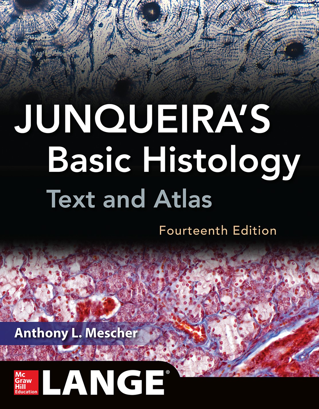
Title: Junqueira's basic histology: text and atlas
Author Name: Anthony L. Mescher.
Author Sur Name: MESCHER, Anthony L.
Author information:
Anthony L. Mescher, PhD is Professor of Anatomy and Cell Biology at the Indiana University School of Medicine Bloomington, Indiana.
Edition/Published: 14th ed. _New York : McGrawHill , 2016
New to this edition:
  1. The blue medical application boxes is one of the most useful aspect of this book. You would find medical application related data in few histology books.
  2. The diagrams and pictures of electron microscope and light microscope that are given with very topic are of very high quality.
  3. All the diagrams of histology are properly labelled.
  4. All the diagrams and images are explained in great detail in the pink boxes. This explanation of different points in diagrams make it one of the most easy to understand histology book.
  5. In the latest edition of junqueira histology all the chapters and revised and text is reduced and newly discovered knowledge is added.
Physical Description:
xii, 560p., : illustrations (chiefly color), tables, diag., chart.; 27.5cm.
Notes Includes Index.
Includes Index: P. 529-560
ISBN No's: 978-1-25-925098-9
Bar Code's:
Shelf Location's: 86
Classification
Subject: Histology
Dewey Class No: 611/.018
Letter Call No: M56j
LC Classification: QL807 .H57 2016
Other's Book Information
Book ID No: 1624
Total Books: 1
Date of collection's: 01-Nov-2016
Donation / Purchase: Purchase
Language: English
Status: Available
Department: Anatomy
Synopsis:
  1. Featuring more than 1000 state-of-the-art photographs and drawings, Junqueira's has been the leading text in medical histology and microscopic anatomy for more than three decades. This 14th edition has been revised to feature a new full-colour presentation, new art, and the addition of an online learning centre.
Review:
  1. This book of basic histology is one of the best resource of histology written by Anthony S. Mescher. This book of histology has 14 editions. And is one of the best selling histology book. Many of the students who want to study histology in depth study this book. So, if you are an undergraduate and want to study histology deeply you should consult this books. Also this is a great resource for those who are postgraduate students of histology or histo-pathology.
Description:
  1. The most authoritative, current, and beautifully illustrated histology text available NEW clinical vignettes have been added to each chapter Full-color, easy-to-understand drawings provide just the right level of detail necessary to reinforce key concepts and facilitate comprehension and retention of text material NEW chapter-ending multiple-choice questions review must-know material For more than three decades, Junqueira's Basic Histology has been unmatched in its ability to explain the function of cell and tissue structure in the human body. Updated to reflect the latest research in the field, and enhanced with more than 1,000 illustrations, most in full-color, the Fourteenth Edition reflects the most comprehensive and modern approach to understanding medical histology available anywhere. This well-regarded classic is distinguished by chapters focusing on the cytoplasmic and nuclear compartment of the cell, the four basic tissues that form the organs, and each organ system. In response to reader demand, the legends are now concise, stand-alone summaries of the illustrations. Applauded for its visual appeal, Junqueira’s is enhanced by full-color micrographs that comprise a complete atlas of tissue sections. These state-of-the-art micrographs highlight the important features of every tissue and organ in the human body, while full-color, easy-to-understand drawings provide just the right level of detail necessary to clarify the text and make learning easier.
Key Features:
  1. Recognized for more than three decades as the most authoritative, comprehensive, and effective approach to understanding medical histology
  2. Unmatched in its ability to explain the relationship between cell and tissues structure and their function in the human body
  3. Updated to the latest research and development on each topic.
  4. New and additional medical applications throughout each chapter provide clinical relevance for every subject
  5. New design incorporates full color for each figure and table in addition to color micrographs of each cell and tissue.
  6. Figure legends summarize and provide easy access to key points of each topic
  7. Author anthony L. Mescher, Ph.D. has over 30 years' experience teaching cell biology and histology to medical students
  8. Summaries of key points at the end of each chapter list main points concisely for very easy review
  9. Many new tables help integrate each histological topic with relevant physiological aspects of the system
  10. New, detailed tables of contents within each chapter
Summary:
Explains the function of cell and tissue structure in the human body. Updated to reflect the research in the field, and enhanced with more than 1,000 illustrations, this title includes chapters focusing on the cytoplasmic and nuclear compartment of the cell, the four basic tissues that form the organs, and each organ system.
Abstract:
This new edition of the best-selling English edition of Junqueira's Basic Histology: Text & Atlas will be available in late 2015. This textbook is written for advanced undergraduate students and medical students seeking a concise yet complete presentation of human microscopic anatomy or histology. Previous editions of this textbook have been translated into over 10 languages and are used in medical colleges worldwide. With the best art program of any histology textbook and the most comprehensive presentation of light and electron micrographs to illustrate all cells and tissues of the human body. The new edition of Junqueira is the best tool yet for gaining a modern understanding of the structural and functional relationships underlying physiology, cell biochemistry, and pathology.
Preface:
With this 14th edition, Junqueira's Basic Histology continues as the preeminent source of concise yet thoroughinformation on human tissue structure and function. For nearly 45 years this educational resource has met the needs of learners for a well-organized and concise presentation of cell biology and histology that integrates the material with that of biochemistry, immunology, endocrinology, and physiology and provides an excellent foundation for subsequent studies in pathology. The text is prepared specifically for students of medicine and other health-related professions, as well as for advanced undergraduate courses in tissue biology. As a result of its value and appeal to students and instructors alike, Junqueira's Basic Histology has been translated into a dozen different languages and is used by medical students throughout the world. This edition now includes with each chapter a set of multiple-choice Self-Test Questions that allow readers to assess their comprehension and knowledge of important material in that chapter. At least a few questions in each set utilize clinical vignettes or cases to provide context for framing the medical relevance of concepts in basic science, as recommended by the US National Board of Medical Examiners. As with the last edition, each chapter also includes a Summary of Key Points designed to guide the students concerning what is clearly important and what is less so. Summary Tables in each chapter organize and condense important information, further facilitating efficient learning. Each chapter has been revised and shortened, while coverage of specific topics has been expanded as needed. Study is facilitated by modern page design. Inserted throughout each chapter are more numerous, short paragraphs that indicate how the information presented can be used medically and which emphasize the foundational relevance of the material learned. The art and other figures are presented in each chapter, with the goal to simplify learning and integration with related material. The McGraw-Hill medical illustrations, now used throughout the text and supplemented by numerous animations in the electronic version of the text, are the most useful, thorough, and attractive of any similar medical textbook. Electron and light micrographs have been replaced throughout the book as needed, and again make up a complete atlas of cell, tissue, and organ structures fully compatible with the students' own collection of glass or digital slides. A virtual microscope with over 150 slides of all human tissues and organs is available: http://medsci.indiana.edu/junqueira/virtual/junqueira.htm. As with the previous edition, the book facilitates learning by its organization: • An opening chapter reviews the histological techniques that allow understanding of cell and tissue structure. • Two chapters then summarize the structural and functional organization of human cell biology, presenting the cytoplasm and nucleus separately. • The next seven chapters cover the four basic tissues that make up our organs: epithelia, connective tissue (and its major sub-types), nervous tissue, and muscle. • Remaining chapters explain the organization and functional significance of these tissues in each of the body's organ systems, closing with up-to-date consideration of cells in the eye and ear. For additional review of what's been learned or to assist rapid assimilation of the material in Junqueira's Basic Histology, McGraw-Hill has published a set of 200 full-color Basic Histology Flash Cards, Anthony Mescher author. Each card includes images of key structures to identify, a summary of important facts about those structures, and a clinical comment. This valuable learning aid is available as a set of actual cards from Amazon.com, or as an app for smart phones or tablets from the online App Store. With its proven strengths and the addition of new features, I am confident that Junqueira's Basic Histology will continue as one of the most valuable and most widely read educational resources in histology. Users are invited to provide feedback to the author with regard to any aspect of the book's features. Anthony L. Mescher Indiana University School of Medicine mescher@indiana.edu
Content:

KEY FEATURES

PREFACE

ACKNOWLEDGMENTS

1 Histology & Its Methods of Study 1

Preparation of Tissues for Study-1

Light Microscopy-4

Electron Microscopy-8

Autoradiography-9

Cell & Tissue Culture-10

Enzyme Histochemistry-10

Visualizing Specific Molecules-10

Interpretation of Structures in Tissue Section-14

Summary of Key Points-15

Assess Your Knowledge-16

2 The Cytoplasm-17

Cell Differentiation-17

The Plasma Membrane-17

Cytoplasmic Organelles-27

The Cytoskeleton-42

Inclusions-47

Summary of Key Points-51

Assess Your Knowledge-52

3 The Nucleus-53

Components of the Nucleus-53

The Cell Cycle-58

Mitosis-61

Stem Cells & Tissue Renewal-65

Meiosis-65

Apoptosis-67

Summary of Key Points-69

Assess Your Knowledge-70

4 Epithelial Tissue-71

Characteristic Features of Epithelial Cells-72

Specializations of the Apical Cell Surface-77

Types of Epithelia-80

Transport Across Epithelia-88

Renewal of Epithelial Cells-88

Summary of Key Points-90

Assess Your Knowledge-93

5 Connective Tissue-96

Cells of Connective Tissue-96

Fibers-103

Ground Substance-111

Types of Connective Tissue-114

Summary of Key Points-119

Assess Your Knowledge-120

6 Adipose Tissue-122

White Adipose Tissue-122

Brown Adipose Tissue-126

Summary of Key Points-127

Assess Your Knowledge-128

7 Cartilage-129

Hyaline Cartilage-129

Elastic Cartilage-133

Fibrocartilage-134

Cartilage Formation, Growth, & Repair-134

Summary of Key Points-136

Assess Your Knowledge-136

8 Bone-138

Bone Cells-138

Bone Matrix-143

Periosteum & Endosteum-143

Types of Bone-143

Osteogenesis-148

Bone Remodeling & Repair-152

Metabolic Role of Bone-153

Joints-155

Summary of Key Points-158

Assess Your Knowledge-159

9 Nerve Tissue & the Nervous System-161

Development of Nerve Tissue-161

Neurons-163

Glial Cells & Neuronal Activity-168

Central Nervous System-175

Peripheral Nervous System-182

Neural Plasticity & Regeneration-187

Summary of Key Points-190

Assess Your Knowledge-191

10 Muscle Tissue-193

Skeletal Muscle-193

Cardiac Muscle-207

Smooth Muscle-208

Regeneration of Muscle Tissue-213

Summary of Key Points-213

Assess Your Knowledge-214

11 The Circulatory System-215

Heart-215

Tissues of the Vascular Wall-219

Vasculature-220

Lymphatic Vascular System-231

Summary of Key Points-235

Assess Your Knowledge-235

12 Blood-237

Composition of Plasma-237

Blood Cells-239

Summary of Key Points-250

Assess Your Knowledge-252

13 Hemopoiesis-254

Stem Cells, Growth Factors, & Differentiation-254

Bone Marrow-255

Maturation of Erythrocytes-258

Maturation of Granulocytes-260

Maturation of Agranulocytes-263

Origin of Platelets-263

Summary of Key Points-265

Assess Your Knowledge-265

14 The Immune System & Lymphoid Organs-267

Innate & Adaptive Immunity-267

Cytokines-269

Antigens & Antibodies-270

Antigen Presentation-271

Cells of Adaptive Immunity-273

Thymus-276

Mucosa-Associated Lymphoid Tissue-281

Lymph Nodes-282

Spleen-286

Summary of Key Points-293

Assess Your Knowledge-294

15 Digestive Tract-295

General Structure of the Digestive Tract-295

Oral Cavity-298

Esophagus-305

Stomach-307

Small Intestine-314

Large Intestine-318

Summary of Key Points-326

Assess Your Knowledge-328

16 Organs Associated with the Digestive Tract-329

Salivary Glands-329

Pancreas-332

Liver-335

Biliary Tract & Gallbladder-345

Summary of Key Points-346

Assess Your Knowledge-348

17 The Respiratory System-349

Nasal Cavities-349

Pharynx-352

Larynx-352

Trachea-354

Bronchial Tree & Lung-354

Lung Vasculature & Nerves-366

Pleural Membranes-368

Respiratory Movements-368

Summary of Key Points-369

Assess Your Knowledge-369

18 Skin-371

Epidermis-372

Dermis-378

Subcutaneous Tissue-381

Sensory Receptors-381

Hair-383

Nails-384

Skin Glands-385

Skin Repair-388

Summary of Key Points-391

Assess Your Knowledge-391

19 The Urinary System-393

Kidneys-393

Blood Circulation-394

Renal Function: Filtration, Secretion, & Reabsorption-395

Ureters, Bladder, & Urethra-406

Summary of Key Points-411

Assess Your Knowledge-412

20 Endocrine Glands-413

Pituitary Gland (Hypophysis)-413

Adrenal Glands-423

Pancreatic Islets-427

Diffuse Neuroendocrine System-429

Thyroid Gland-429

Parathyroid Glands-432

Pineal Gland-434

Summary of Key Points-437

Assess Your Knowledge-437

21 The Male Reproductive

System-439

Testes-439

Intratesticular Ducts-449

Excretory Genital Ducts-450

Accessory Glands-451

Penis-456

Summary of Key Points-457

Assess Your Knowledge-459

22 The Female Reproductive System-460

Ovaries-460

Uterine Tubes-470

Major Events of Fertilization-471

Uterus-471

Embryonic Implantation, Decidua, & the Placenta-478

Cervix-482

Vagina-483

External Genitalia-483

Mammary Glands-483

Summary of Key Points-488

Assess Your Knowledge-489

23 The Eye & Ear: Special Sense Organs-490

Eyes: The Photoreceptor System-490

Ears: The Vestibuloauditory System-509

Summary of Key Points-522

Assess Your Knowledge-522

APPENDIX-525

FIGURE CREDITS-527

INDEX-529

Read