Anwer Khan Modern Medical College
Library Management System
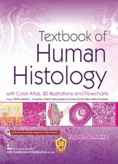
| Title: | Textbook of human histology: with color atlas, 3D illustrations and flowcharts. |
| Author Name: | Yogesh Ashok Sontakke |
| Author Sur Name: | SONTAKKE, Yogesh Ashok. |
| Author information: |
|
| Edition/Published: | 1st ed. _New Delhi : CBS Publishers , 2020 |
| New to this edition: |
|
|
Physical Description: xii, 346, : 27.5 cm |
| Notes | Includes bibliographical references and indexes. |
| Includes Index: | 335-346 |
| ISBN No's: | 978-81-94125-42-6 |
| Bar Code's: | |
| Shelf Location's: |
| Classification | |
| Subject: | Histology |
| Dewey Class No: | 611 |
| LC Classification: | QM551 .S54 2020 |
| Other's Book Information | |
| Book ID No: | 2669 |
| Total Books: | 1 |
| Date of collection's: | 18-Mar-2023 |
| Donation / Purchase: | Donation |
| Language: | English |
| Status: | Available |
| Department: | Anatomy |
| Synopsis: |
|
| Description: |
|
| Key Features: |
|
| Summary: |
The "Textbook of human histology" is a comprehensive guide to the microscopic anatomy of human tissues and organs. It provides detailed descriptions of the structure and function of cells, tissues, and organs, and includes color atlas illustrations, 3D illustrations, and flowcharts to aid in understanding. The book covers a wide range of topics, including the basics of cell biology, the structure and function of epithelial, connective, muscular, and nervous tissues, and the structure and function of specific organs such as the heart, lungs, liver, and kidneys. The book also includes clinical correlations that relate histological findings to clinical conditions and diseases, making it a valuable resource for medical students, healthcare professionals, and researchers. Overall, the book provides a comprehensive and detailed overview of human histology, and the inclusion of color atlas illustrations, 3D illustrations, and flowcharts make it a helpful resource for visual learners. |
| Abstract: |
The book is a comprehensive textbook on human histology with an emphasis on visual aids such as a color atlas, 3D illustrations, and flowcharts. It is authored by Yogesh Sontakke, published by CBS Publications, and was first released on May 30, 2019. It has received positive feedback and is targeted toward medical and health science students, as well as professionals in related fields. |
| Preface: |
This Textbook of Human Histology has been written to fulfill the requirements of students and teachers as per the Competency Based Undergraduate Curriculum for Indian Medical Graduates.* As per the new curriculum, histology learning involves • Knowledge (K) and skill (S) learning domains • Knows (K) and knows how (KH) levels of competencies as per Miller’s pyramid# Most of the histology topics are at the core level of competency (must-know). Only a few topics are in noncore levels of competency (nice to know/desirable to know) such as cardio esophageal junction, corpus luteum, olfactory epithelium, eyelid, lip, sclerocorneal junction, optic nerve, cochlea, organ of Corti, pineal gland, and ultrastructure of muscular tissue, nervous tissue, epithelium, and connective tissue. The histology-related competencies should be taught with lectures and practical classes. Assessment methods for histology competencies involve written examination and skill assessment to identify the given slide with interpretation. Various topics of histology need horizontal integration with physiology and vertical integration with pathology. To fulfill above requirements, this textbook is provided with the following features: • Concise text with functional correlation for quick recapitulation during examination (horizontal integration). • 117 Photomicrographs help to identify the microscopic structures. • 122 Flowcharts help to revise and memorize the microanatomy. • 106 Practice figures (H&E pencil drawings) are easy to draw for written assessments. • 175 Three-dimensional illustrations provide a visual grasp and easier retention of difficult concepts. • Summary (examination guide) to overcome the difficulty of summarizing the facts in written assessments. • Identification feature markings focus readers on specific points (for knows, knows how levels of competencies). • Neet, MCQ, Viva voce, and Clinical fact markings for preparation of various upcoming academic entrance examinations. • Tables to summarize essential facts. • Boxes to focus on important topics. • Some interesting facts to isolate them from the main text, so that these facts should not be missed. • Clinical correlation oriented toward the pathogenesis of diseases (vertical integration). Students are suggested to read the book in the following sequence: Text ® Flowchart ® Summary (examination guide) ® draw practice figures in the log book Any suggestions from the reader for rectification and improvement are welcome.
Yogesh Ashok Sontakke dryogeshas@rediffmail.com |
Related Books
- Langman's medical embryology.
- Atlas of human anatomy
- Atlas of human anatomy
- Clinical anatomy by regions
- Gray's anatomy: the anatomical basis of clinical practice
- Junqueira's basic histology: text and atlas
- Textbook of anatomy upper limb and thorax: vol. 1
- Textbook of anatomy head, neck and brain: vol. 3
- Textbook of human osteology: with atlas of muscle attachments
- Textbook of human osteology: with atlas of muscle attachments