Anwer Khan Modern Medical College

Library Management System
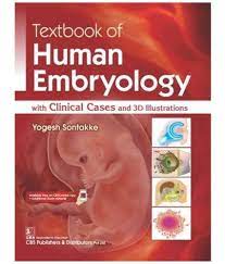
Advance Search

Book Detail

Title: Textbook of human embryology: with clinical cases and 3D lllustrations
Author Name: Yogesh Ashok Sontakke
Author Sur Name: SONTAKKE, Yogesh Ashok.
Author information:
<p>Yogesh Sontakke MBBS, MD (Anatomy) is currently Associate Professor, Department of Anatomy, Jawaharlal Institute of Postgraduate Medical Education and Research (JIPMER), Pondicherry, an institution of national importance under the Ministry of Health and Family Welfare, Government of India. He is also author of the books: Principles of Clinical Genetics, Principles of Histological Techniques, Immunohistochemistry and Microscopy, and Principles of Scientific Writing. Dr.  He has authored several books including Principles of Scientific Writing, Textbook of Human Embryology with Clinical Cases And 3D Illustrations, Principles of Histological techniques, Immunohistochemistry and Microscopy, and Principles of Clinical Genetics.</p> <p>Dr. Sontakke's work has contributed to the advancement of medical knowledge and education in India and helped medical students and professionals gain a deeper understanding of anatomy and its applications in clinical practice. His contributions to the field of anatomy may have also been recognized and celebrated within the Indian medical community.</p> <p> </p>
Edition/Published: 1st ed. _New Delhi : CBS Publishers , 2019
New to this edition:
  1. -New or updated information on the latest research in embryology.

    -Updates on the use of new technologies or techniques in the study of human embryology.

    -Clinical case studies that reflect recent developments in medical practice or treatment options.

    -Additional 3D illustrations or other visual aids to help students better understand the complex processes of human embryology.

    -Discussion of new ethical or legal considerations related to embryology or reproductive technology.

Physical Description:
viii, 320p., : 27.8cm
Includes Index: 315-320
ISBN No's: 978-93-88108-37-9
Bar Code's:
Shelf Location's:
Classification
Subject: Embryology
Dewey Class No: 612.6
LC Classification: RG629
Other's Book Information
Book ID No: 2670
Total Books: 1
Date of collection's: 18-Mar-2023
Donation / Purchase: Donation
Language: English
Status: Available
Department: Anatomy
Synopsis:
  1. Textbook of Human Embryology: With Clinical Cases and 3D Illustrations” is a concise textbook that has been written in easy-to-understand language for ease of exam revision. The book has 3D illustrations that provide images of embryological changes, while flowcharts make the revision of developmental sequences easier. Tables summarise all the essential facts and boxes focus on the most important topics students need to remember to of Form

Description:
  1. The "Textbook of Human Embryology: With Clinical Cases and 3D Illustrations" by Dr. Yogesh Ashok Sontakke is a comprehensive guide to human embryology, providing a detailed exploration of the different stages of embryonic development. The book features clear explanations of embryological concepts and processes, accompanied by numerous clinical cases and 3D illustrations that bring the subject matter to life.

    The book is divided into seven chapters, starting with an introduction to embryology and the stages of human embryonic development. It then covers gametogenesis and fertilization, early development, the development of specific systems and organs, the placenta and fetal membranes, teratology, and applied embryology. Throughout the book, there are detailed descriptions of the structure and function of different organs and systems, as well as the mechanisms involved in their development.

    The "Textbook of Human Embryology" is an essential resource for medical students, researchers, and healthcare professionals seeking a comprehensive understanding of human embryology. The inclusion of clinical cases and 3D illustrations make the subject matter accessible and engaging, while the clear explanations and detailed descriptions ensure that readers gain a solid grounding in the fundamental principles of embryology.

Key Features:
  1. “Textbook of Human Embryology: With Clinical Cases and 3D Illustrations” is an exceptional textbook on embryology with 3-dimensional illustrations drawn to create a life-like effect in understanding the intricacies of developing human structure. The book focuses on clinical cases with clinical facts. With concise text, the highlights of the book include flowcharts, tables, summary (examination guide), boxes, interesting facts, electron micrographs, clinical photographs, and imaging studies.

Summary:

"Textbook of Human Embryology: With Clinical Cases and 3D Illustrations" is a comprehensive textbook that provides a detailed overview of the development of the human embryo from fertilization to birth.

The book is authored by Dr. Yogesh Ashok Sontakke, a renowned Indian medical author and anatomist, and includes clinical cases and 3D illustrations to help students understand the complex processes of embryology.

Some of the key topics covered in the book include:

  • The early development of the embryo, including fertilization, cleavage, and gastrulation.
  • The formation and development of the various organ systems, including the nervous system, cardiovascular system, and digestive system.
  • The process of fetal development, including fetal circulation, growth, and the development of various organs.
  • Clinical cases and case studies that demonstrate the application of embryological concepts to real-world medical situations.
  • 3D illustrations and other visual aids that help students visualize the complex processes of embryology.

Overall, "Textbook of Human Embryology: With Clinical Cases and 3D Illustrations" is a valuable resource for medical students, anatomists, and anyone else interested in understanding the complex processes of human embryology.

Abstract:

The book is a concise textbook that has been written in easy-to-understand language for ease of exam revision. The book has 3D illustrations that provide images of embryological changes, while flowcharts make the revision of developmental sequences easier. Tables summarise all the essential facts and boxes focus on the most important topics students need to remember

Preface:

This Textbook of Human Embryology has been written keeping in mind the requirements of students and teachers. Due to the complexity of the subject, readers face difficulty in understanding and imagining the developing human structures. Hence, in the present book, an attempt has been made to provide all necessary information for easy understanding.
User-friendly features of this book with their purposes are as follows:
• Concise text is given in easy language; unnecessary details are avoided.
• 3D illustrations to provide easy imagining of developing structures.
• Flowcharts to revise and memorize the developmental sequence.
• Tables to summarise essential facts.
• Summary (examination guide) to overcome the difficulty of summarising the facts in theory examinations.
• Neet, MCQ, Viva Voce and Clinical facts markings for preparation of various upcoming academic entrance examinations.
• Boxes to focus on important topics.
• Interesting facts to isolate them from the main text, so that these facts should not be missed by readers.
• 40 Scanning electron micrographs to give real insight into developing structures.
• 70 Clinical cases for early clinical exposure of various clinical anomalies encountered by eminent clinicians.
• Special topics such as assisted reproductive techniques (including in vitro fertilization) and ultrasonography in embryology are also included to give orientation toward clinical aspects.
I am hopeful that this book will help to fulfill all the requirements of students and teachers. Students are suggested to read in the following sequence:
Illustrations → Stages of development → Flowchart →
Table → Summary (examination guide) → Practice
Figure
Any suggestions from the readers for rectification and improvement are welcome at dryogeshas@rediffmail.com

Content:

1. Introduction-1
2. Gametogenesis-6
3. Menstrual Cycle-16
4. First Week of Development-24
5. Assisted Reproduction Technology: In vitro Fertilisation and Intracytoplasmic Sperm Injection-34
6. Second Week of Development-40
7. Third Week of Development-49
8. Embryonic Period: Four to Eight Weeks of Development-61
9. Placenta and Umbilical Cord-73
10. Integumentary System: Skin, its Appendages and Mammary Gland-86
11. Pharyngeal Apparatus-96
12. Alimentary Tract I: Development of Face, Nose, Palate-109
13. Alimentary Tract II: Development of Teeth, Pharynx, Tongue and Salivary Glands-117
14. Alimentary Tract III: Development of Intestine-126
15. Alimentary Tract IV: Development of Liver, Gallbladder, Pancreas, and Spleen-141
16. Respiratory System-149
17. Development of Body Cavities and Diaphragm-158
18. Cardiovascular System I: Development of Heart-168
19. Cardiovascular System II: Blood Vessels and Foetal Circulation-184
20. Urinary System: Kidney, Ureter, Urinary Bladder, Urethra-203
21. Reproductive System: Male and Female Reproductive Organs-215
22. Nervous System-231
23. Development of Eye-257
24. Development of Ear-266
25. Endocrine System-273
26. Skeletal System and Limbs-279
27. Muscular System-293
28. Foetal Period: Nine Weeks to Birth-297
29. Clinical Applications and Ultrasonography in Embryology-300
30. Multiple Pregnancy (Twinning)-305
Annexures-310
I. Embryonic Remnants-310
II. Placenta Previa-311
III. Hermaphrodite-312
IV. Derivatives of Neural Crest Cells-313
Index-315

Read